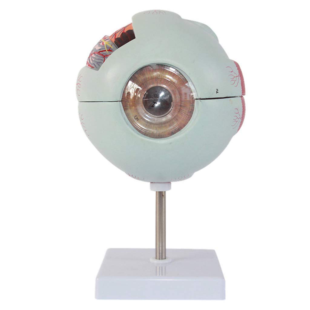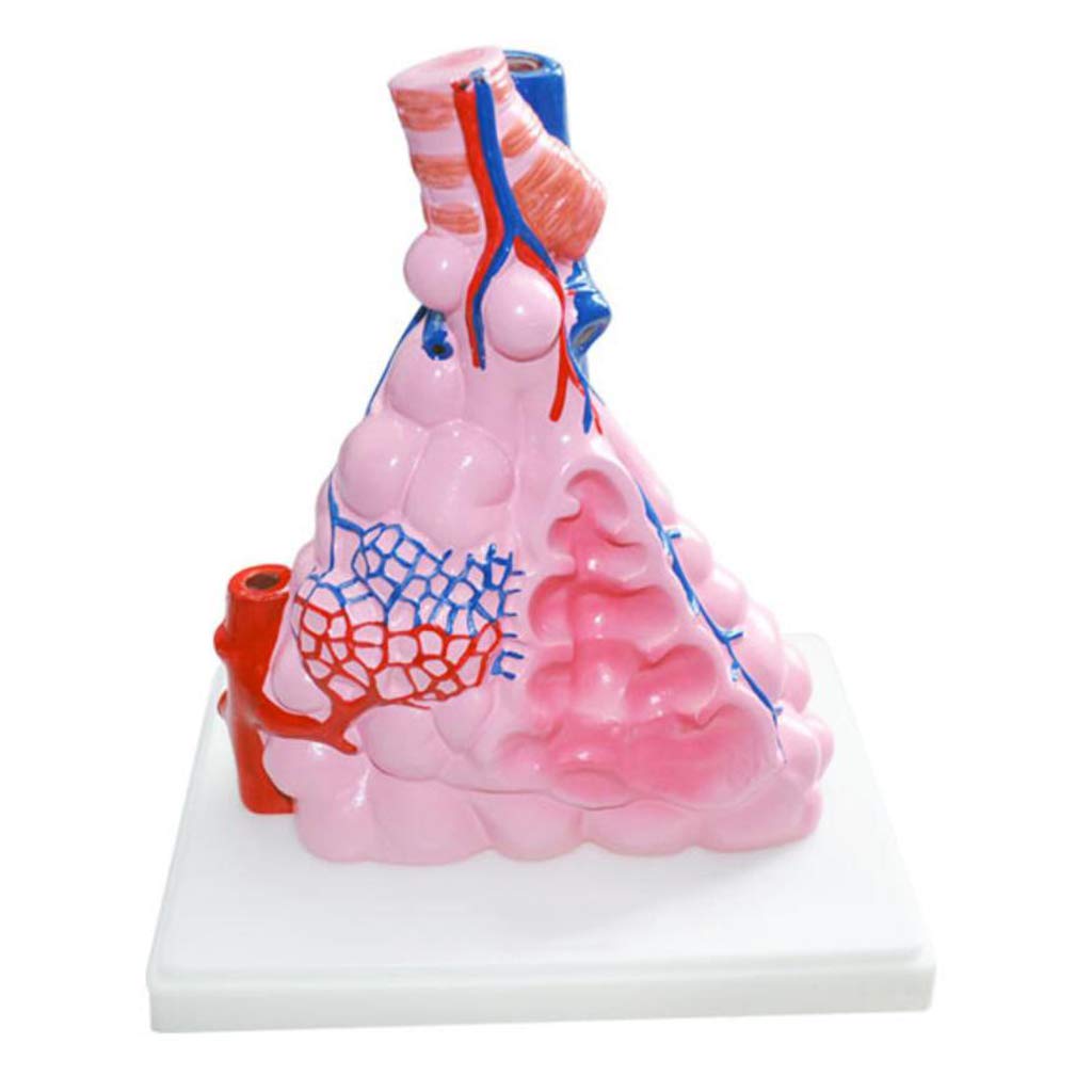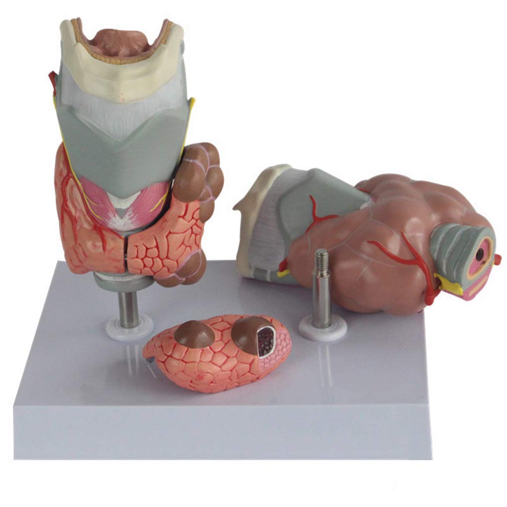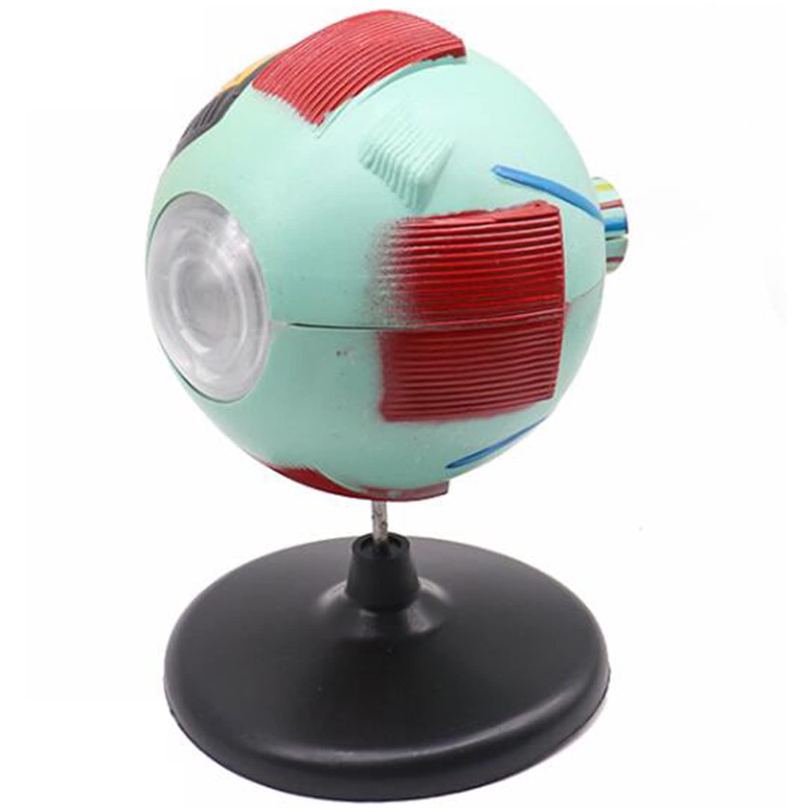
Sjdopulse 6 Times Magnified Eyeball Model Medical Science Human Eyes Anatomy Teaching Model Advanced Human Eyeball Model For Primary And Secondary Schools Lab Supplies
sjdoPulse
Teaching Content: Perform horizontal cross section through the front and rear pole of the eyeball. The cross section of the ball wall shows three outer layers, middle and inner layers. The outer layer shows: the cornea, sclerosis and cornea are transparent and occupy 1/6 of the eyeball surface. Professional: this model consists of human eye samples, reliable textbooks and atlases and is 6 times larger than the eyeball Suitable for physiology and hygiene teaching in high school and anatomy teaching in medical schools, medical schools, nursing schools and health schools. Show details: The curvature of the front is larger than the hardeners, and the back curve is larger than the front. On the front edge of the film you can see the venous sinus of the hardera. The middle layer shows: retina, macular, visual disc, retina, arteries, also visible the front border of the retina: teeth, edge. Biology Teaching Tools - Different colors are used to distinguish different positions, and the colors are bright and easily attract the attention of students, so that you can learn demonstration, which promotes understanding of students and increases classroom fun. Material: made of food-grade environmentally friendly PVC materials, high quality prototype, handmade in sturdy, undamaged condition. If you have any questions, please email me, I will reply you within 24 hours
Compare prices (1 shop)
| shop | Price | Action |
|---|---|---|

|
41,75 GBP | Go to shop |